
News Articles
New imaging technology for Radiographers
Health and Environmental Sciences
Clinical Sciences
Teaching and Learning
CUT News
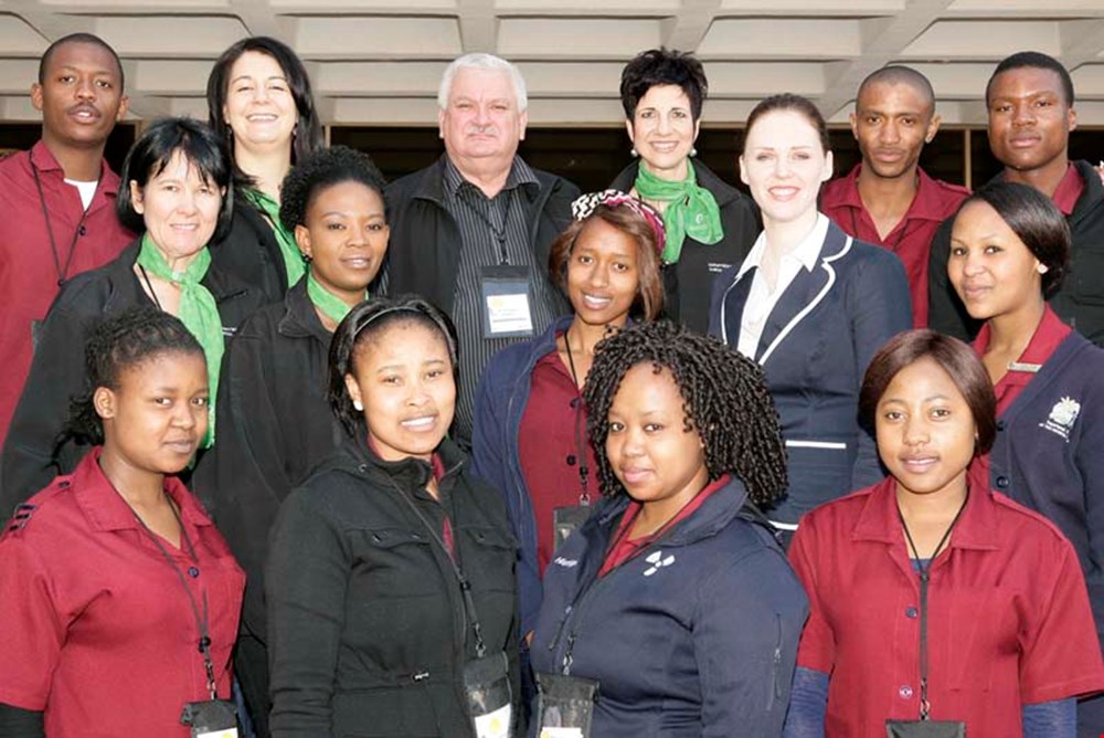
Department of Clinical Sciences in the Faculty of Health and Environmental Sciences at CUT held a two-day annual seminar for Radiographers, Radiologists, and Technical Engineers on 15 and 16 August 2014.
The purpose of the event is to ensure that stakeholders and hospital staff where CUT students are placed for their Work-Integrated Learning (WIL), get an opportunity to obtain continuous development, exposure to the latest trends and technology and networking. This year, a special session showed radiographers the history and the links between x-ray imaging, ultrasound and nuclear medicine. Not only was the new technology links shared with the audience but families involved in creating the medical equipment were at the seminar! A new imaging technology called capsule endoscopy was also explored (see image above). The capsule is the size and shape of a pill and contains a tiny camera. After a patient swallows the capsule, it takes pictures of the inside of the gastrointestinal tract. The presenters also shared with the audience, information regarding the competition commission, protection of private information, how to determine bone age, and the best practice for shoulder pathology. Apart from the medical topics discussed, the theme that was explored and exploited was “What is the verdict?”. The intention of the theme was to raise awareness of professional behaviour in the workplace, labour law, and the overall occupation relationship between employer and employee.
Image(s)
Pictured is a group of delegates who attended the two day engagement.
News


